
UCL acadmics produce winning image in the science photo competition Wellcome Image Awards 2015
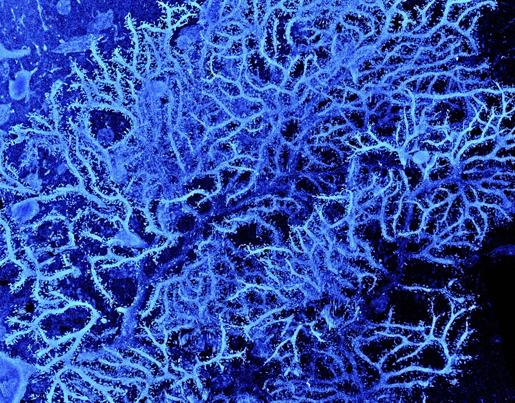
A scanning electron micrograph of tree-like branches (dendritic tree) spreading out from a particular type of nerve cell (Purkinje cell, or neurone) found in the brain. The finger-like projections in this elaborate network act like tiny sensors, picking up information and passing on messages to help control and coordinate muscle movement. This particular neurone is from the cerebellar cortex in a rat brain. To allow us to see the dendritic tree, this Purkinje cell was filled with a visual marker before being imaged by focused ion beam scanning electron microscopy, which allows neurones and neural circuits to be reconstructed in high resolution. The width of the image is 110 micrometres (0.11 mm)
What is focused ion beam scanning electron microscopy?
Scanning electron microscopy creates detailed images using a focused beam of high-energy electrons to scan the surface of a sample or specimen. Electrons are slowed down when they come into contact with a solid sample, and this produces a variety of signals (including secondary and back-scattered electrons, X-rays, light, and heat) that can be detected. Some of these signals are used to create detailed images of the surface texture of the sample. Images produced in this way are greyscale because electrons don’t contain information about colour, but false colour can be digitally added to an image to highlight particular areas of interest or to create visual impact.
Focused ion beam scanning electron microscopy also uses a beam of electrons to create images. In addition to this, there is a second beam made up of gallium ions that comes into contact with the sample at the same point as the electron beam. This gallium ion beam can be used in a similar way to a laser to cut into the sample, for example to produce very thin slices. By coupling this with a traditional electron beam, these can then be imaged to produce scanning electron micrographs. By stacking these slices back together in the right order, a high-resolution 3D image of the whole specimen can be obtained.
Click here for information on the facilities at the LCN including the Focussed Ion Beam Microscope.
Related articles are in the BBC news, the Guardian and the Telegraph.