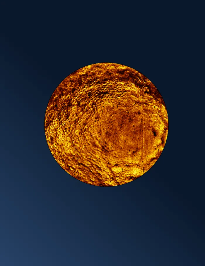
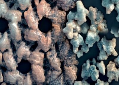
The study, published in the journal Nature Communications, used a new technique to keep live bacteria in place so the cells could be imaged without wriggling away.
The method, in which bacteria were chemically attached to a glass surface, was so effective that, if left for a longer period (about 50 minutes), many bacteria remained standing straight up from the glass they were immobilised on, instead of lying down on their sides. This allowed researchers to image the upward facing ends.
The bacteria were imaged using atomic force microscopy, where a tiny probe only a few nanometres wide gently taps across the bacterial cell like a finger reading Braille, “feeling” the surface to create an image at a much higher resolution than is possible using light microscopy.
By better understanding the structure of bacterial surfaces, the researchers aim to lay the foundations for new approaches to target deadly drug-resistant strains.
“Imaging the poles of bacteria is like opening a view of the back of the moon. We are looking at something that no one else has seen before. The poles are where cells divide and so contain critical information about this process. They may also be a particular target for antibiotics, with some bacteria-killing treatments preferring to attack this part of the cell.
“Our method of immobilising the cells without affecting their ability to grow and reproduce also means we can image a much larger range of live bacteria than was previously possible. This is important as it may help us understand differences between species and why antibiotics work for some of them but not others.
“Our work is fundamental science that ultimately aims to find new leads to target drug-resistant bacteria.”
Senior author Professor Bart Hoogenboom (London Centre for Nanotechnology at UCL and UCL Physics and Astronomy)
“Our immobilisation method opens up exciting new possibilities. We can now study the armour of bacteria across different species more reliably and clearly observe nanoscale differences that were previously difficult to assess.”
Lead author Dr Anna Scheeder, based at the London Centre for Nanotechnology at UCL, said:
The team, which included researchers from the University of Oxford, coated a glass surface with a chemical that bound to proteins on the bacterial surface, keeping the cells in place without affecting their viability. Cells were immobilised horizontally after 20 minutes; left a little longer, for 50 minutes, and many of them were unexpectedly found to stand straight up.
The researchers found the poles of more active bacteria (i.e., ones that were dividing rapidly) had fold-like defects compared to less active bacteria that had more smoothly rounded ends, hinting that this part of the cell might not have fully remodelled after division.

Pole of a stationary (i.e., not rapidly dividing) E. coli bacterium, left, compared to a more active E. coli cell, right. Credit: Anna Scheeder / Bart Hoogenboom.
As well as imaging the poles of E. coli (Escherichia coli) bacteria, the researchers found striking differences between E. coli and two other bacterial species, Pseudomonas aeruginosas and Acinetobacter baumannii, which, like E. coli, are known for their ability to evade common antibiotics and cause difficult-to-treat infections.
The two strains, which have never been imaged at this resolution before, lacked the network of little holes (pores) found in the surface of E. coli, suggesting their response to antibiotics, as well as their intrinsic resistance mechanisms, may differ significantly.
The work was funded by the Wellcome Trust, the Biotechnology and Biological Sciences Research Council (BBSRC), the Rosetrees Trust, and the Engineering and Physical Sciences Research Council (EPSRC).